39 draw and label the stages of a nematocyst discharge
Unit 2 Objectives - Cnidaria Flashcards | Quizlet Study with Quizlet and memorize flashcards containing terms like Explain the General Characteristics shared by most Cnidarians (include 2 body forms), List and briefly explain the animals that belong to the 3 classes of Cnidaria, Draw, label, and explain the structures of Hydrozoa and more. Nematocysts - The Stinging Cells | Zoology for IAS, IFoS and other ... The discharge of nematocyst thread tube from the cnidocyte takes place due to mechanical or chemical stimuli received by the cnidocil. In a resting stage the capsule wall is not permeable to water and there is very high osmotic pressure since the hypnotoxin inside is hypertonic to external water.
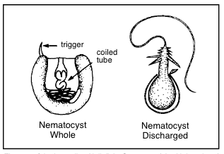
Formation and discharge of nematocysts is controlled by a proton ... A nematocyst consists of a capsule containing a coiled tubule. On triggering, the cyst extrudes this tubule in an extremely rapid manner. The mechanisms and driving forces of discharge are still unknown.

Draw and label the stages of a nematocyst discharge
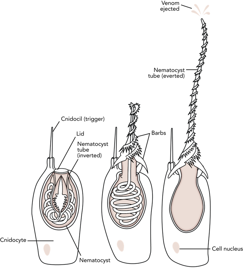
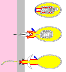
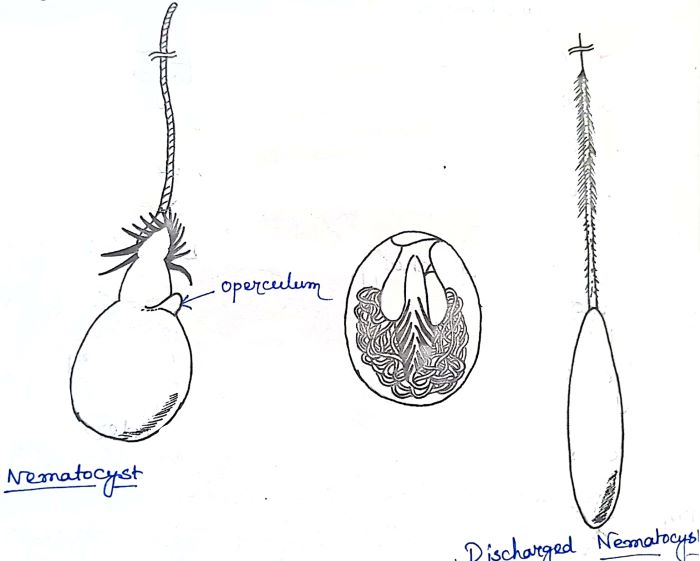
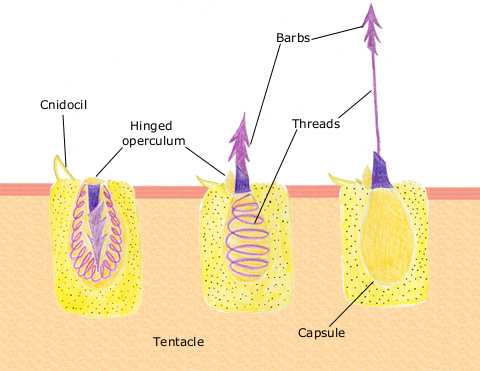
The cnidarian nematocyst: a miniature extracellular matrix ... - PubMed Nematocyst discharge, which is one of the fastest processes in biology, is driven by an extreme osmotic pressure of about 150 bar. The molecular analysis of the nematocyst has from the beginning indicated a collagenous nature of the capsule structure. Stages Of Nematocyst Discharge - Lara Sheppard Discharge mechanism of a nematocyst. A nematocyst consists of a capsule containing a coiled tubule. Cnidaria, representing the first stage of metazoan evolution, . Nematocyte discharge results from stimulation of the cnidocil ciliary cone. It shows the nematocyst in three stages of discharge. Phylum Cnidaria - SlideShare The capsule is capped at its outer margin by an operculum that is discharge of the nematocyst. The trigger like cnidocil is resposible for nematocyst discharge. (b) A discharge nematocyst. When the cnidocil is stimulated a rapid (osmotic0 influx of water causes the nematocyst to evert, first near its base, and then progressively along the tube ...
Draw and label the stages of a nematocyst discharge. Nanosecond-scale kinetics of nematocyst discharge - Current Biology The rapid discharge of stinging cells (nematocytes) in jellyfish, hydra, and other cnidarians is one of the fastest movements in the animal kingdom. After the triggering of exocytosis, a miniature cellular weapon, the nematocyst, is released and stylets punch a hole into the prey's integument. This step is so fast that conventional high-speed micro-cinematography fails to resolve its kinetics [1]. Nematocyst Stingers Accelerate Fast — Biological Strategy - AskNature The Strategy. The cells (cnidocysts) produce one large organelle called a nematocyst. The cell forms a layered matrix around the nematocyst that keeps it strong and promotes the generation of 150 bar of pressure within the organelle at maturation. The "lid" of the capsule (operculum) associates with the cell membrane facing out. Nematocysts - WikEM Management. Remove tentacles and nematocysts. Hot salt water immersion (inactivates heat labile toxins), submerging the area in 45 ℃ water for 20 minutes has been shown to provide significant pain relief [2] Stingose is a topical solution composed of 20% aluminum sulfate and 1.1% surfactant. Used for pain control, venom removal via osmosis ... Activity: Nematocysts | manoa.hawaii.edu/ExploringOurFluidEarth Label the cnidocil, lid, cell nucleus, nematocyst tube and barbs where applicable. Test the response of the nematocysts to a hair root Pull out a head hair with a root. Insert the root of the hair under the coverslip, touching the tentacle tis¬sue while your partner watches through the microscope. Pull the root slightly away from the tentacle.
How Do Jellyfish Sting? - Ocean Conservancy Nematocyst stay safely in their sacs when the animal is unbothered. But when the animal senses either a physical or chemical disturbance, such as an unsuspecting beach-goer bumping into a jellyfish tentacle, the nematocyst will shoot out of the sac. The entire discharge takes about three milliseconds—faster than you can get away. Discharge Mechanism of the Nematocysts of Pelagia noctiluca Abstract. The nematocysts are complex organelles, contained in specialized cells, the nematocytes. Under adequate stimulus (Lubbock 1979; Tardent et al. 1980; Ertman and Davenport 1981) the nematocysts eject a threat that, in turn, either adheres to or penetrates into the prey, injecting the venomous substances contained into the capsule fluid. Nematocyst | biology | Britannica As eversion and twisting proceed, the barbs act like a drill, penetrating into (and pulling the thread into) the foreign object. If a toxin is present, it passes through the hollow thread, penetrating and paralyzing the victim's tissues. After eversion, the thread separates from the nematocyst. Cnidocyte - Wikipedia Discharge mechanism of a nematocyst The cnidocyst capsule stores a large concentration of calcium ions, which are released from the capsule into the cytoplasm of the cnidocyte when the trigger is activated. This causes a large concentration gradient of calcium across the cnidocyte plasma membrane.
Jellyfish Nematocysts | Uncommon Descent Nematocysts (also known as cnidocysts) of jellyfish and other cnidarians are giant exocytotic organelles of the stinging cells used for prey capture and defense. These miniature cellular weapons contain a cocktail of hemolytic and neurotoxic poisons, making some cnidarians the most venomous animals known. Injection of the toxins requires an ... 3D Printed Nematocyst | Creature Cast | Learn Science at Scitable It shows the nematocyst in three stages of discharge. First, the inverted harpoon is packed within the capsule. The harpoon is inside-out at this point. Second, a stimulus has led the capsule to ... PDF Cnidarians: Jellyfish, coral, hydra & sea anemones Cnidarian Basics Label the cross-section of the two forms of Cnidarians: Flashy Features: ... 30.Where are the 2 main locations of nematocysts? a. b. 31.What causes a Nematocyst to discharge? 32.How often can the mechanism in the nematocyst be triggered? 33. Draw and label the stages of a Nematocyst discharge. Human Uses: Micrographs of discharged nematocyst. The major discharged nematocysts ... Micrographs of discharged nematocyst. The major discharged nematocysts in this study are shown. A and B were from A. aurita. C, D, and E were from C. pacifica. F and G were from C. yamaguchii. H...
Nematocyst discharge in Polykrikos. (A) The putative ...
Draw And Label The Stages Of A Nematocyst Discharge Tika sumpter southside screening attend legend chicago john. Draw and label the stages of a nematocyst discharge.Serita jakes on twitter: ""you are an #amazingdad😇 a # John legend, tika sumpter attend 'southside with you' chicago screening Ayusya home health care pvt ltd-bangalore-chennai-madurai-coimbatore Diablo succubus wretched succubi wiki wikia

A) Four stages of the discharge of a Hydra stenotele hitting ...
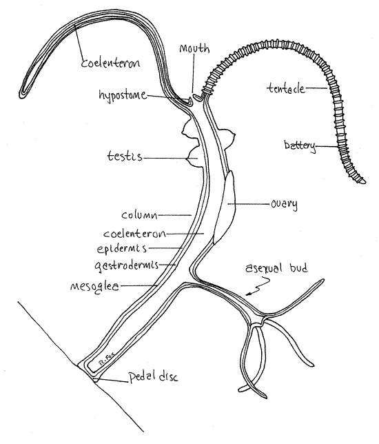
Porifera and Cnidaria Flashcards | Quizlet Nematocysts are a specialized cell in the tentacles of a cnidarian containing a barbed or venomous coiled thread that can be projected in self-defense or to catch prey. List 4 functions of nematocysts. -Locomotive -Defensive -Paralyzing -Grasping Where are the 2 main locations of nematocysts? -Ectoderm and Endoderm -Tentacles

Elastodiagnosis of diseases: A review - ScienceDirect
Phylum Cnidaria - SlideShare The capsule is capped at its outer margin by an operculum that is discharge of the nematocyst. The trigger like cnidocil is resposible for nematocyst discharge. (b) A discharge nematocyst. When the cnidocil is stimulated a rapid (osmotic0 influx of water causes the nematocyst to evert, first near its base, and then progressively along the tube ...
Phylum Cnidaria | manoa.hawaii.edu/ExploringOurFluidEarth
Stages Of Nematocyst Discharge - Lara Sheppard Discharge mechanism of a nematocyst. A nematocyst consists of a capsule containing a coiled tubule. Cnidaria, representing the first stage of metazoan evolution, . Nematocyte discharge results from stimulation of the cnidocil ciliary cone. It shows the nematocyst in three stages of discharge.

The regulation of cnidocyte discharge - ScienceDirect
The cnidarian nematocyst: a miniature extracellular matrix ... - PubMed Nematocyst discharge, which is one of the fastest processes in biology, is driven by an extreme osmotic pressure of about 150 bar. The molecular analysis of the nematocyst has from the beginning indicated a collagenous nature of the capsule structure.

Zoology Chapter 7 Flashcards | Quizlet

animal cnidaria Flashcards | Quizlet

Figure 1 from Ultrastructure of the dinoflagellate Polykrikos ...
Frontiers | Expression of Opsins of the Box Jellyfish ...

Cnidocyte - Wikiwand

30+ Hydra Anatomy Illustrations, Royalty-Free Vector Graphics ...

A) Four stages of the discharge of a Hydra stenotele hitting ...
Hydra (genus) - Wikipedia

A) Four stages of the discharge of a Hydra stenotele hitting ...
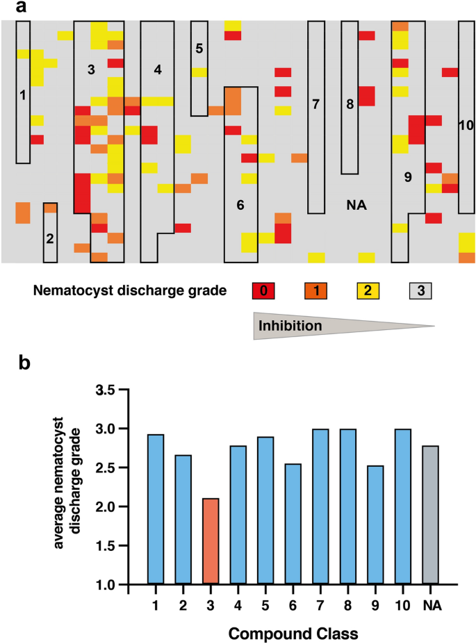
A small molecule screen identifies novel inhibitors of ...

407 Cnidocytes Images, Stock Photos & Vectors | Shutterstock

Cnidocyte | everyEthing
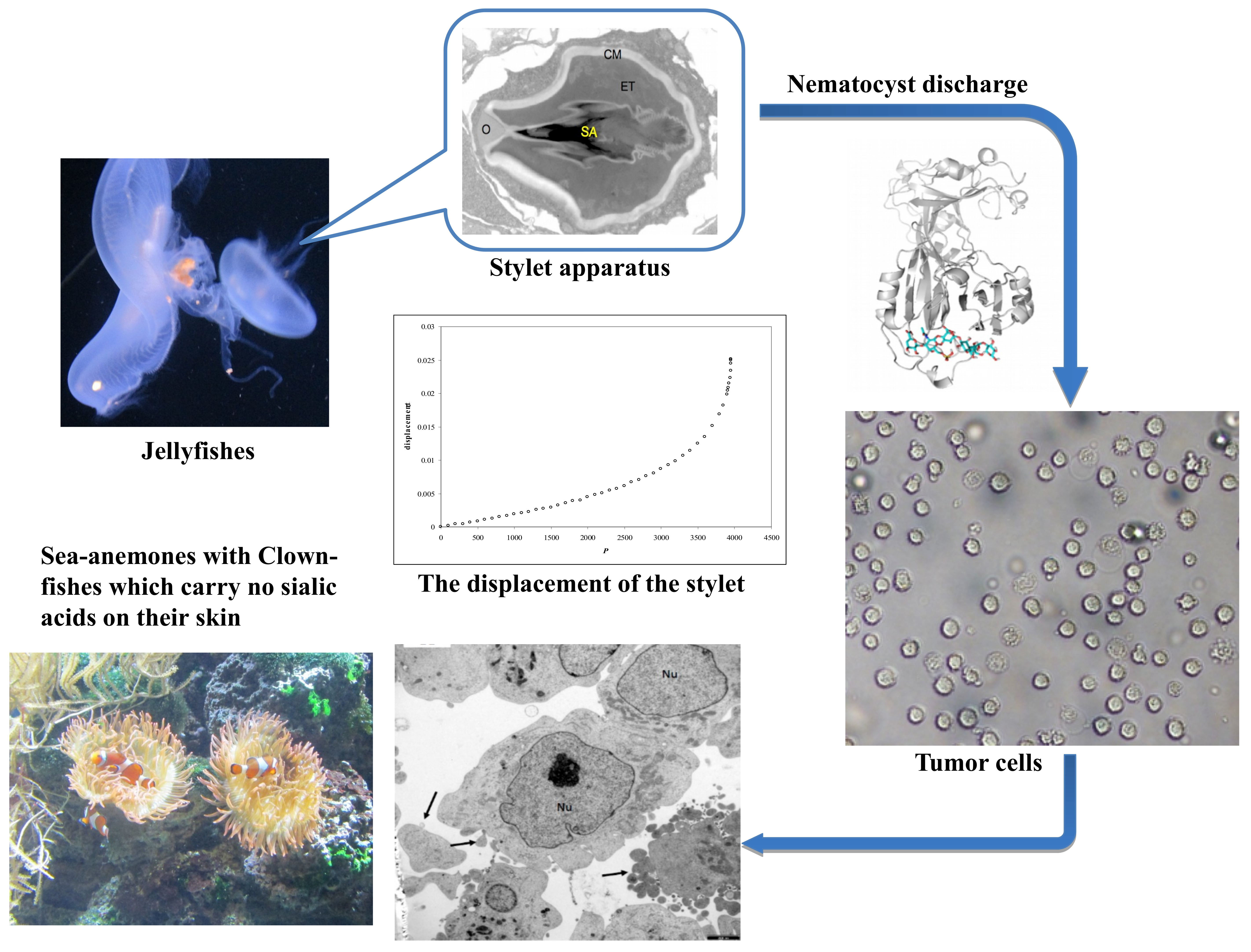
Marine Drugs | Free Full-Text | The Sialic Acid-Dependent ...

Model of elastic nematocyst discharge. (A) schematic ...
Non-muscle myosin II drives critical steps of nematocyst ...

Expression profiling and cellular localization of myxozoan ...

The nematocyst: a molecular map of the cnidarian stinging ...

Figure 3 from Dart formation in nematocysts of the sea ...
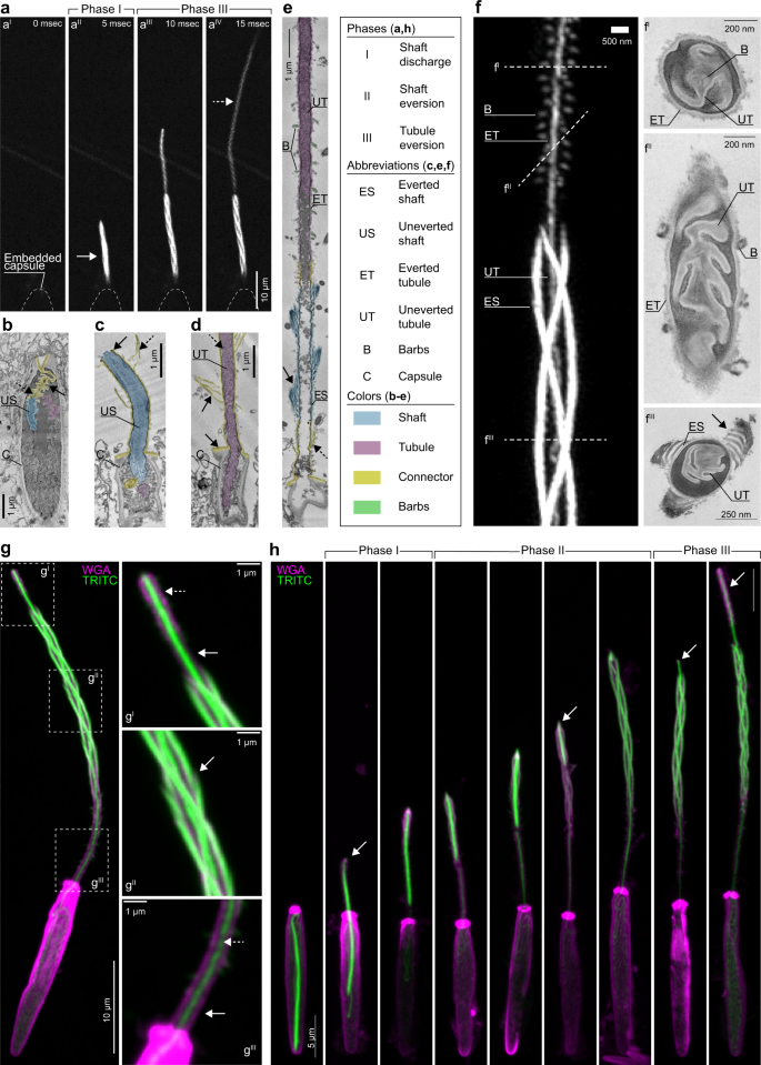
The architecture and operating mechanism of a cnidarian ...
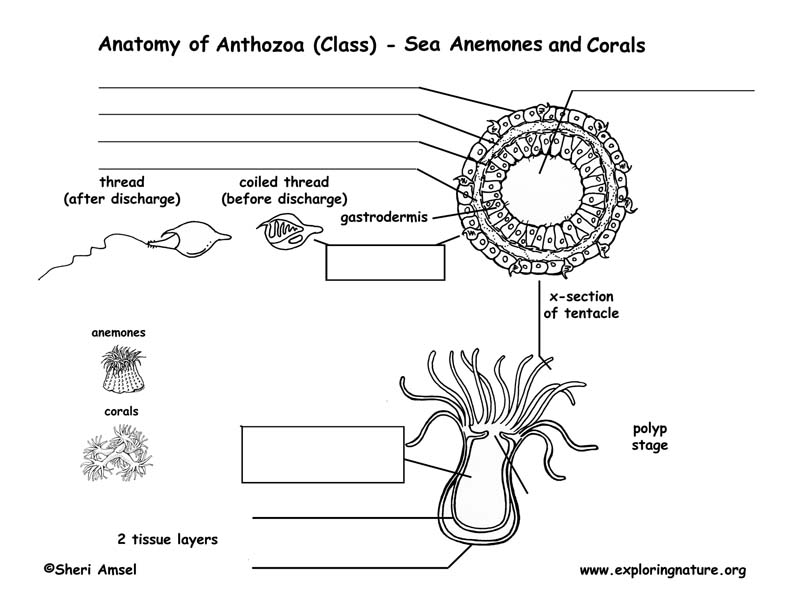
Class - Anthozoa (Sea Anemones, Corals)
Experimental Investigation on the Diffusion Law of Polymer ...

Phylum Cnidaraia (COELENTERATA) (knide, nettle) | Biology Boom
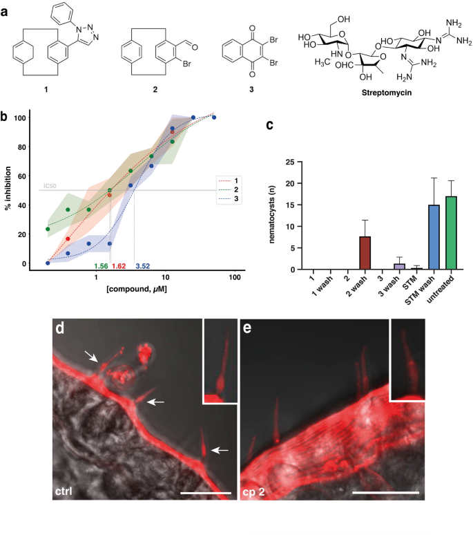
A small molecule screen identifies novel inhibitors of ...
Global Diversity and Review of Siphonophorae (Cnidaria ...
Untitled 1
Cnidocyte | everyEthing
Jellyfish in Alabama - Alabama Cooperative Extension System

Structure and function of a typical Cnidaria nematocyst. a ...

Chapter 7 Diagram | Quizlet

Phylum Cnidaria | Biology for Majors II

File:Nematocyst-discharge process.png - Wikipedia
File:Nematocyst discharge.png - Wikimedia Commons
Cnidome and Morphological Features of Pelagia noctiluca ...
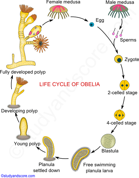
Obelia - Structure, Diagram, Life Cycle | Solved Question
Post a Comment for "39 draw and label the stages of a nematocyst discharge"